Visualizing and analyzing trajectories
Overview
Teaching: 20 min
Exercises: 10 minQuestions
How to load structure files and trajectories into VMD?
How to animate trajectories?
How to generate RMSD data from a trajectory?
Objectives
Learn how to load molecular structure files and MD trajectories.
Learn how to animate trajectories?
Get workshop example data
On the training cluster:
cp /project/60104/workshop_vmd_2024.tar.gz .
tar -xf workshop_vmd_2024.tar.gz
On any other computer:
wget https://github.com/ComputeCanada/molmodsim-amber-md-lesson/releases/download/workshop-2021-04/workshop_vmd_2024.tar.gz
Loading trajectory files
A trajectory file contains the coordinates for all atoms over the course of a simulation. Normally, not every time step of the simulation is saved, since it would create a huge file. It is common to save coordinates every 1000 to 5000 steps. All of these coordinates allow us to measure the dynamic properties of our simulation experiments, including secondary-structure evolution, diffusion constants, correlations between groups, etc
Loading structure files
To load a trajectory, we need both the structure and the trajectory file.
First load a structure file as a new molecule. VMD can read structure files in different formats, such as AMBER7 Parm, XPLOR PSF, GROMACS GRO, PDB, etc.). File types are recognized by extension. If a file has a non-standard extension you can select format manually.
It is best to use structure files that contain connectivity information whenever possible. Examples of file formats with connectivity are molecular topology files or mol2 files. In the absence of connectivity information, VMD uses distances between atoms to determine which ones are connected. Automatic bond determinations does not work perfectly all the time. Stretched bonds may go undetected, and there may be incorrect bonds formed between non-bonded atoms are too close to each other. If you use the wrong bonds, your visualization will be incorrect. The automatic bond determination can be disabled when loading structure files from the command line.
- To load a trajectory, we need both the structure and the trajectory file.
- It is best to use structure files that contain connectivity information whenever possible.
As an example, open Tk Console and run the following commands to load the file 7xcq.pdb without automatic bond determination:
cd ~/workshop_vmd/example_01
mol new 7xcq.pdb autobonds off
Delete all the molecules or restart vmd.
Our training simulation dataset is located in the directory workshop_vmd/example_02. As a structure file we will be using AMBER7 parameter file prmtop_nowat.parm7. Change into this directory and load the topology file.
Once a molecular structure has been loaded you can add a trajectory to it: highlight the molecule, go to File->Load Data into Molecule and choose mdcrd_nowat.xtc. It is a long trajectory with 3000 frames. To make loading faster load every 5th frame.
Loading trajectory using commands on the training cluster
cd ~/workshop_vmd/example_02
mol new prmtop_nowat.parm7
mol addfile mdcrd_nowat.xtc step 5
Viewing AMBER-NetCDF trajectories on Windows and MAC.
NetCDF trajectory files, which are AMBER’s default format, can only be loaded on Linux. You can convert them to GROMACS XTC format by using the CPPTRAJ program from AMBER.
module load ambertools
cpptraj prmtop_nowat.parm7
trajin mdcrd_nowat.nc trajout mdcrd_nowat.xtc go
Visualizing trajectories
- To make trajectory animation run smoother you can interpolate coordinates:
Graphical representations->Trajectory->Trajectory Smoothing Window Size - You can also visualize periodic images:
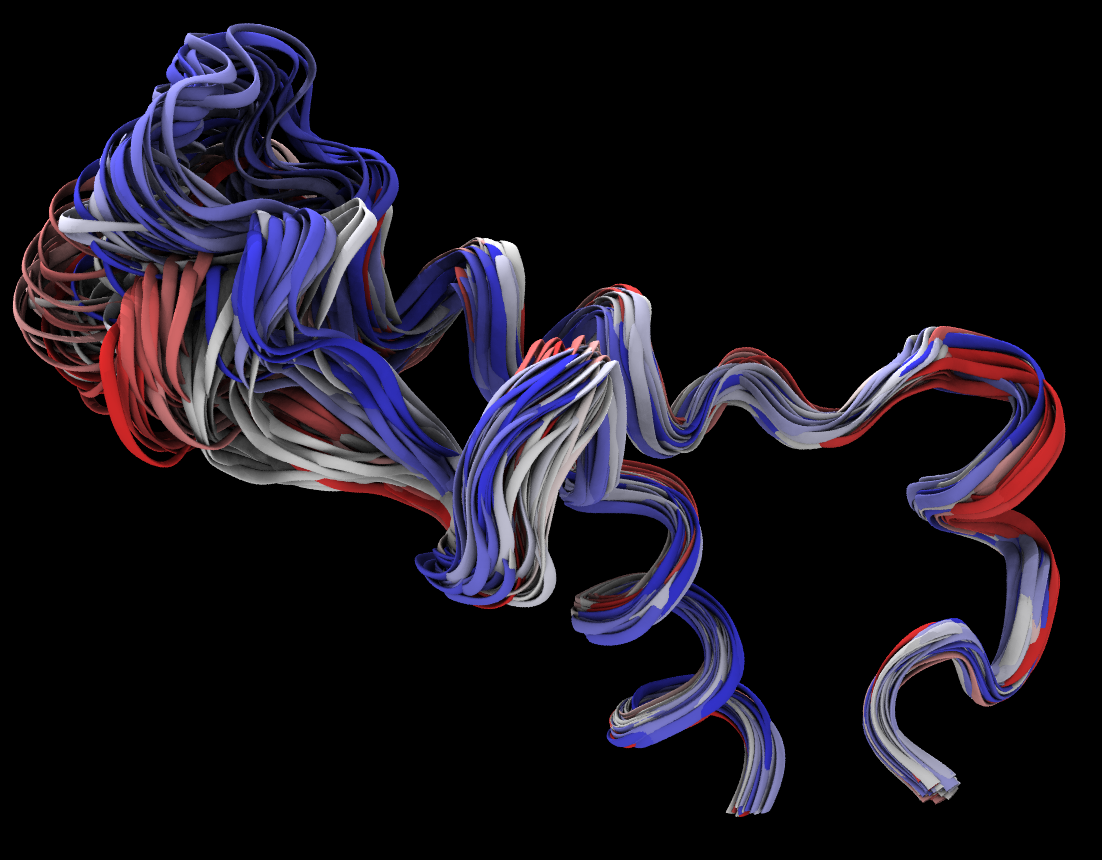
Graphical representations->Periodic - You can visualize multiple frames and color them by trajectory step. Try resid 809 to 859.
RMSD Trajectory Analysis
The RMSD is a numerical measurement of the difference between two structures: a target structure and a reference structure. Our interest in molecular dynamics is in how structures and parts of structures change over time. For example, a plot of RMSD vs. time will reveal the opening and closing of gates on a protein, such as a transporter. When compared with the starting point, the RMSD can identify protein structure changes and study stability of the simulated system. As the RMSD curve flattens or levels off, that can be a sign that the system has equilibrated.
- You can calculate the time dependence of RMSD in a molecular dynamics simulation using the RMSD Trajectory Tool. It is located under
Extensions->Analysis->RMSD Trajectory Tool
- Load a trajectory
- Start
RMSD Trajectory Tooland add a molecule:Add active Alignall frames using backbone of the whole protein. You can choose a reference frame or a reference molecule.- Make a selection of atoms for which you want to calculate RMSD
- use all protein backbone atoms (normally you don’t want to include hydrogens)
- use all nucleic acid atoms
- use a specific part ot the system (e.g. resid 20 to 80)
- You can optionally save rmsd in a file so you can make a nice figure with your favorite plotting software, and check
Plotbox to view the result.
Calculate the RMSD for two groups of atoms over time
Align frames using backbone of all protein residues. Compute trajectory RMSD for two selections of backbone atoms: residues 790-810 and 820-840.
- Considering both selections, what is the minimum and the maximum RMSD?
- How does the RMSD change when you include all atoms?
- Over the course of the simulation, which of the groups is more stable?
- Are your RMSD results affected by the previous superposition step?
Solution
- The minimum is 0.39, the maximum is 6.1.
- RMSD increases when all atoms are considered.
- The first group, 790-810.
- Yes
RMSD Calculator
The RMSD calculator is similar to the RMSD Trajectory Tool, but it calculates the RMSD between two molecules. It is located under Extensions->Analysis->RMSD Calculator.
Calculation of the RMSD between two molecules
The RMSD calculator works well when two molecules are composed of the same atoms, but the alignment will fail if atom selection in the reference molecule differs from that in the target molecule. The issue is illustrated in this exercise.
- Compute RMSD of two molecules: PDB ID 1si4 and 4n7n. For the calculation, use only chain A backbone atoms.
- When all chain A residues are used for alignment, why does the alignment fail?
- Can you think of a way to include all backbone atoms present in both proteins in the alignment?
If you need to download pdb files use:
wget https://files.rcsb.org/download/4n7n.pdbSolution
- Use the atom selection:
chain A and resid 1 to 140, and check boxBackbone onlyfor both alignment and RMSD calculation, RMSD = 0.92558- The residue 141 of 1si4 molecule has the terminal oxygen atom “OXT”, while it is absent in 4n7n.
- Exclude the OXT atom from the selection:
not name OXT and chain A and resid 1 to 141
Key Points